We performed a quantitative depth-resolution analysis of two twisted hexagonal boron nitride (h-BN) flakes reconstructed from an experimental multislice electron ptychography dataset [1]. The depth resolution was determined from the buried heterointerfaces, where atomic columns disappear in one flake and appear in the other. The results are in good agreement with the theoretical depth-resolution equation based on the maximum diffraction angle [2].
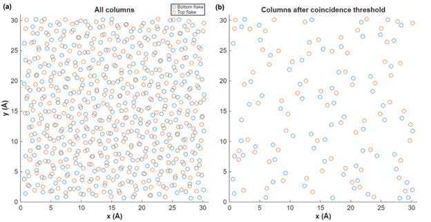
We first identified the atomic column centers in the top and bottom flakes at the buried interface and retained only those columns that satisfied a positional coincidence criterion of 0.9 Å, that is, the minimum distance between two columns is 0.9 Å. Using the retained columns, we extracted depth profiles by averaging the reconstructed phase-sum signal within an in-plane cylinder of 0.25 Å radius centered on each column. Each depth profile was normalized and treated as an edge spread function (ESF), from which we quantified the depth resolution using the standard 10–90% ESF width [3]. This edge-response metric is conservative in practice because any deviation from an ideal step transition, including finite interface width, residual roughness, and reconstruction smoothing, broadens the measured 10–90% transition. In that case, the measured ESF width overestimates the true axial blur, meaning the true depth resolution can be better than the reported value.
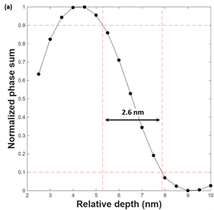
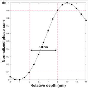
For the top flake, we analyzed falling edges at the buried interface. The best column yields a depth resolution of 2.6 nm. Averaging over the 10 sharpest columns gives 3.0 ± 0.2 nm, and averaging over the 50 sharpest columns gives 3.5 ± 0.3 nm. Varying the cylinder radius to 0.35 Å and 0.5 Å yields consistent results. For the bottom flake, we analyzed rising edges at the buried interface. The best column yields 3.0 nm. Averaging over the 10 sharpest columns gives 3.2 ± 0.1 nm, and averaging over the 50 sharpest columns gives 3.5 ± 0.2 nm. Again, changing the cylinder radius to 0.35 Å and 0.5 Å produces very similar values.
We next compare the experimental results with the theoretical depth-resolution equation [2],

where λ is the electron wavelength and θ is the maximum diffraction angle. Using an electron energy of 300 keV and θ = 36.2 mrad in our experiment, Eq. (1) gives ddep = 3.0 nm. Experimentally, averaging the 10 sharpest columns from the top flake and the 10 sharpest columns from the bottom flake yields an average depth resolution of 3.1 nm, in close agreement with the 3.0 nm prediction. In contrast, a conventional aperture-based depth-of-focus estimate using a probe convergence semi-angle α = 17.1 mrad gives an aperture-limited depth scale of about 6.7 nm using the standard λ/α2 scaling. This is worse than the experimentally measured 2.6 to 3.5 nm range obtained from the multislice ptychography reconstruction, consistent with the fact that the reconstruction leverages higher-angle scattering information up to θ = 36.2 mrad rather than being limited by the probe convergence semi-angle alone.
In conclusion, by restricting the analysis to buried-interface columns and using the standard 10–90% ESF criterion, we obtain a depth resolution of 2.6–3.5 nm. Averaging the 10 sharpest columns from the top flake and the 10 sharpest columns from the bottom flake yields an average depth resolution of 3.1 nm. This experimental result is in good agreement with Eq. (1), supporting the depth-resolution estimate reported for the experimental multislice electron ptychography reconstruction. All data and analysis codes, including this file, are available on Zenodo [4] for independent download and testing.