Atomic Electron Tomography (AET)
Addressing Feynman’s 1959 challenge
In his famous "There's plenty of room at the bottom" lecture in 1959, Richard Feynman challenged the physical science community saying “It would be very easy to make an analysis of any complicated chemical substance; all one would have to do would be to look at it and see where the atoms are… I put this out as a challenge: Is there no way to make the electron microscope more powerful?” More than fifth years later, we performed a sequence of groundbreaking experiments to fully address Feynman’s challenge (1). Using a novel tomographic reconstruction method, known as equal slope tomography (EST) that we developed in 2005 (2), we demonstrated for the first time electron tomography at 2.4 Å resolution using Au nanoparticle in 2012 (3). Individual atoms were observed inside the nanoparticle and several 3D grains were identified at atomic scale resolution. After this pioneering work, we applied this 3D imaging method to observe nearly all the atoms in a Pt nanoparticle. Atomic steps at 3D twin boundaries were revealed and the 3D core structure of edge and screw dislocations were imaged at atomic resolution (4). These dislocations and the atomic steps at the twin boundaries, which appear to be stress-relief mechanisms, were not visible in conventional 2D projections.
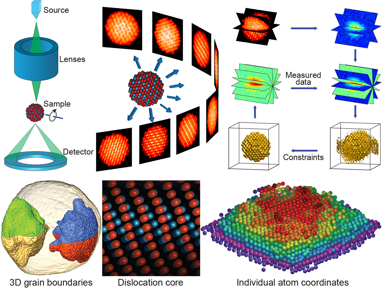
More recently, we reported, for the first time, the determination of the 3D coordinates of thousands of individual atoms and a point defect in a material with a precision of ~19 picometer, where the crystallinity of the sample was not assumed (5). From the coordinates of these individual atoms, we measured the atomic displacement field and the full strain tensor with a 3D resolution of 1 nm and a precision of 10-3, which were further verified by density functional theory calculations. The ability to precisely localize the 3D coordinates of individual atoms in materials without assuming crystallinity, identify point defects in three dimensions, and measure the atomic displacement field and the full strain tensor is expected to transform our understanding of materials properties and functionality at the most fundamental level (1).
Selected Publications
1. J. Miao, P. Ercius and S. J. L. Billinge, "Atomic electron tomography: 3D structures without crystals", Science 353, aaf2157 (2016).
2. J. Miao, F. Förster and O. Levi, "Equally Sloped Tomography with Oversampling Reconstruction", Phys. Rev. B. 72, 052103 (2005).
3. M. C. Scott, C. C. Chen, M. Mecklenburg, C. Zhu, R. Xu, P. Ercius, U. Dahmen, B. C. Regan and J. Miao, "Electron tomography at 2.4-ångström resolution", Nature 483, 444–447 (2012).
4. C. C. Chen, C. Zhu, E. R. White, C.-Y. Chiu, M. C. Scott, B. C. Regan, L. D. Marks, Y. Huang and J. Miao, "Three-dimensional imaging of dislocations in a nanoparticle at atomic resolution", Nature 496, 74–77 (2013).
5. R. Xu, C.-C. Chen, L. Wu, M. C. Scott, W. Theis, C. Ophus, M. Bartels, Y. Yang, H. Ramezani-Dakhel, M. R. Sawaya, H. Heinz, L. D. Marks, P. Ercius and J. Miao, "Three-Dimensional Coordinates of Individual Atoms in Materials Revealed by Electron Tomography", Nature Mater. 14, 1099-1103 (2015).